- This is a X-ray image of a patient with an implanted pacemaker.
- Source :Link
- To start with let us define pace maker as an electronic device that artificially makes the heart to beat. the heart has its own natural pacemaker. when it fails to work an artificial pacemaker is implanted. In the figure, you can see the image of the pacemaker removed from a deceased patient before cremation.

Where why and when is it used? How does it work? How can a human body work on electric current? In order to understand these questions we first have to understand the electrical system in human body.
Electrical signals in the human body
You are moving your hand. Your brain sends commands to the muscles in your hand to move and hence your hand obeys. What is this command? How does it travel so quickly? It is because of electrical charges. Let us look in detail.
Inside the human body, electric charges are present. Without these electric charges a human cannot live. Any organ eye, nose, etc cannot function without electric charges or flow of electric charges (electric current). How are these electric charges conducted throughout the body or in simple terms how does the flow of these electric charges occur? The nerve cells conduct these electric charges. Because of which we can think, remember, and make decision, act, react and so on. If we speak, listen and watch it is because of those electrical signals. These signals are called action potential.
Our body is made up of millions and millions of cells. Every cell has various constituents in them like proteins (made of an acid called amino acid), carbohydrates (made of carbon, hydrogen). But, a common characteristic of every human cell is as follows. The inside of the cell is always negatively charged and outside the cell is positively charged. The charges are separated by the cell wall. This potential that is present on the boundary of every cell wall is called resting potential. The positive charges are because of sodium and potassium cations. The following figure depicts resting potential

There is always a balanced state towards which all human cells work i.e. to ensure more positive ions remain outside the cell. In other words they make sure they always have resting potential. When you touch a pen, it is your brain feeling it. Hence, there is a messenger carrying this signal to the brain that you have touched a pen and your brain acts accordingly lift it or start writing. As your fingers touch the pen, there is a change in the charges in those cells that come in contact to the surfaces of your finger. The positive ions move inside and the potential changes and this potential is called action potential and now the cell is said to be depolarized.

Because of induction of charges the adjacent cells also undergoes the changes as shown in the figure below. Likewise, the depolarization happens throughout the nerve cells that connect your finger and the brain. As this action potential reaches the brain or when the brain cells get depolarized they recognize it is a pen and send commands to the fingers.

This is how electrical signals travel in the body. There are charges present in the heart too. Electrical charges in the heart conducts in the same way but not through nerves. Let us look in detail.
Electrical system in the heart
To understand the electrical system in the heart let us discuss the working of the heart in brief. The heart is a muscle that works continuously like a pump.Heart is located at the centre of the chest tilted lightly towards left hand side. It is hollow and is divided into four chambers namely
- Left atrium
- Right atrium
- Left ventricle
- Right ventricle
as shown in the figure below. Each beat of the heart is set in motion by an electrical signal generated inside the heart all by itself. In order to understand the electrical system of the heart let us discuss the
 working of the heart in brief
Working of the heart
The right atrium receives impure blood (de-oxygenated blood) from all parts of the body and the left atrium receives pure blood (oxygenated blood) from the lungs. When the atria are full with blood, they contract and hence a pressure is generated. The right atrium contracts first and then the left atrium. Hence there is a time delay between the two atrial contraction. This pressure is called systolic pressure and forces the valve between the atria and ventricles to open and hence the blood flows into ventricles. When the ventricles are full with blood, the ventricles contract.
The pressure developed due to the contraction of ventricles is called diastolic pressure. The right ventricle pumps impure blood to the lungs and the left ventricle pumps pure blood to different parts of the body.In the figure above the impure blood vessels are red in colour and pure blood vessels are blue in colour. From the working of the heart, it is clear that the blood is pumped because of the pressure created due to the contraction of the atria and ventricles. How do the contraction occur? It is because of the electrical signals in the heart, the contraction is happening. These electrical signals originate in a node called sino-atrial node also called as SA node as shown in the figure below. You can see the SA node in the right atrium.

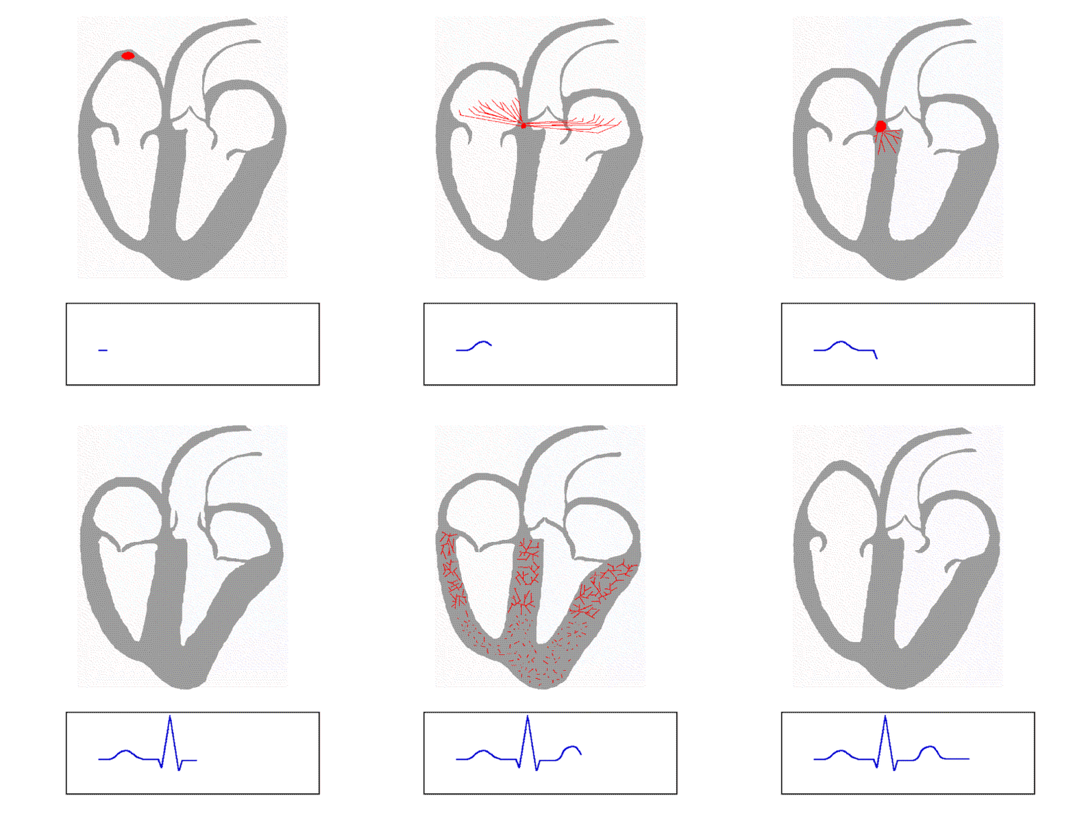
When a cell that is negatively charged becomes positively charged it is called depolarization as already discussed. Depolarization in the heart begins at the Sino-Atrial node also known as SA node. SA node cells can depolarize all by themselves and this action is called automacity .Once the SA node cells are depolarized, the neighboring muscle cells start getting depolarized shown by red arrow around the SA node. This wave of depolarization now spreads through Bachmann's bundle to the left atrium. The atrial contraction happens because of the depolarization of the cells. The cells in the heart are called myocytes and have this special characteristic feature to get reduced in volume when they are depolarized. This happens because of the movement of calcium, potassium and sodium ions inside and outside the cell thereby decreasing and increasing its charge. And correspondingly, the volume changes and hence the contraction and expansion of the atria which leads to filling of blood inside the atria and then being pushed into the ventricles.
Recollect that there is a time delay between the left and right atrial contraction. This is because the electrical signals are generated in the SA node in the right atrium. It takes time for these electrical signals to travel to the left atrium through Bachmann's bundle. Hence the right atrium contracts first and then the left atrium. These signals also travel to another node called Atrio-Ventricular node also called as AV node from the SA node through the path called inter nodal track as shown in the figure above. The distance the signals have to travel from the SA node to AV node is more than the distance they have to travel from SA node to the left atrium. Hence the contraction of ventricles happen after the left atrial contraction.
From the AV node, the signals travel to the walls of the ventricles through bundle of his as shown in the figure. Also, the right ventricle receives the signals first and then the left ventricle. Hence the contraction of right ventricle happens first and then the left ventricle. Hence the order of the contraction of the heart muscle or the order in which electrical signals are received by the heart muscles is as follows
- Right atrium
- Left atrium
- Right ventricle
- Left ventricle
The ECG Electro-cardio Gram, is the recording of the measure of these electrical signal. If you observe an ECG, there are four main waves that are due to the depolarization of the atria and ventricles.

sinus rhythm label by Agateller (Anthony Atkielski) | Link
Here, the waves are the measure of the electrical action in the heart
P wave - depolarization of right atrium
R wave - depolarization of right ventricle
T wave - re-polarization of right ventricle
The PR interval is very important in designing the pacemaker and is the delay between the atria and ventricle depolarization and is 0.2 sec.
The following animation depicts the working of the electrical system in the heart

Now that we have understood the generation and conduction of electrical signals in the heart let us now give pacemaker a proper definition and understand its working. Heart rate is the number of heart beats per minute. When the human body is at rest, the heart rate in adults is 60-80 beats per minute. In other words the SA node generates electrical signal to depolarize the cells 60-80 times in a minute. Hence every cell in the heart depolarizes 60-80 times in a minute. A normal healthy heart has its own pacemaker, the SA node that generates the electrical signals for the heart to pump blood. The heart is in need of a pacemaker under following conditions
Bradycardia – a condition in which the heart beats too slowly - less than 60 beats per minute
Tachycardia - a condition in which the heart beats too fast - more than 80 beats per minute
Atrial fibrillation – the upper chambers of the heart beat rapidly
Heart failure – a condition in which the heartbeat is not sufficient to supply a normal volume of blood
When the above conditions are left untreated, the patient may eventually die. hence to treat the condition or disease, an artificial electronic device is surgically implanted to regulate the heart beat. Such an electronic device is called pacemaker. On the whole, a pacemaker is required if the entire electrical system or a part of the system fails.
What is a pacemaker?
A pace maker is a small device that is implanted under the skin, most often under the collar-bone on the left or right side of the chest. Pacemaker continuously monitors the heart and if it detects a slow rhythm or an interrupted rhythm it sends electrical impulses to make the heart beat normal. As shown in the first figure pacemaker have thin, soft, insulated wires called leads to carry the electrical impulses from the pacemaker to the heart. Pacemaker is designed to mimic the heart's natural pacemaker, the SA node. Pacemaker has two purposes namely
- Pacing
- Sensing
The most popularly used pacemaker nowadays is the dual chamber pacemaker. A dual-chamber pacemaker typically requires two pacing leads: one placed in the right atrium, and the other placed in the right ventricle. A dual-chamber pacemaker monitors (senses) electrical activity in the atrium and/or the ventricle to see if pacing is needed. When pacing is needed, the pacing pulses of the atrium and/or ventricle are timed so that they mimic the heart’s natural way of pumping. It comprises of three parts: an electrical pulse generator, a power source (battery) and an electrode (lead) system. The electrical pulse generator consists of the following components: a sense amplifier circuit, a timing control circuit and an output driver circuit (electrical impulse former) as shown in the figure below

The electrodes give the electrical activity of the heart. After the sense amplifier amplifies the signal, a comparator compares the signals received from the electrodes to the one already stored in the form of voltage signal. If the signal received is the same as that of the reference voltage, then the heart is normal and no signals are sent. But if it is low or high, then the pulse generator starts sending signals till the heart's activity returns to normal. A timing control is needed to control the time difference between the pulses to the SA node and AV node which is 0.2 sec.
This is a very basic working of the pacemaker. In the modern era, the pacemakers with microprocessors and transceivers are under research. These modern pacemakers fit into the application called tele-monitoring of patients where the patient monitoring system is linked to the satellites and the doctors can get updates of their patients' condition from any part of the world. Such pacemakers record the history of working of the heart and its electrical activity and transmits data when in need.

No comments:
Post a Comment